Arch Aortogram
Anatomy normal arch aortic aorta illustration radiopaedia continuation version size full ascending Arch aortogram showing transection of the aorta just distal to the left Arch aortic right heart congenital
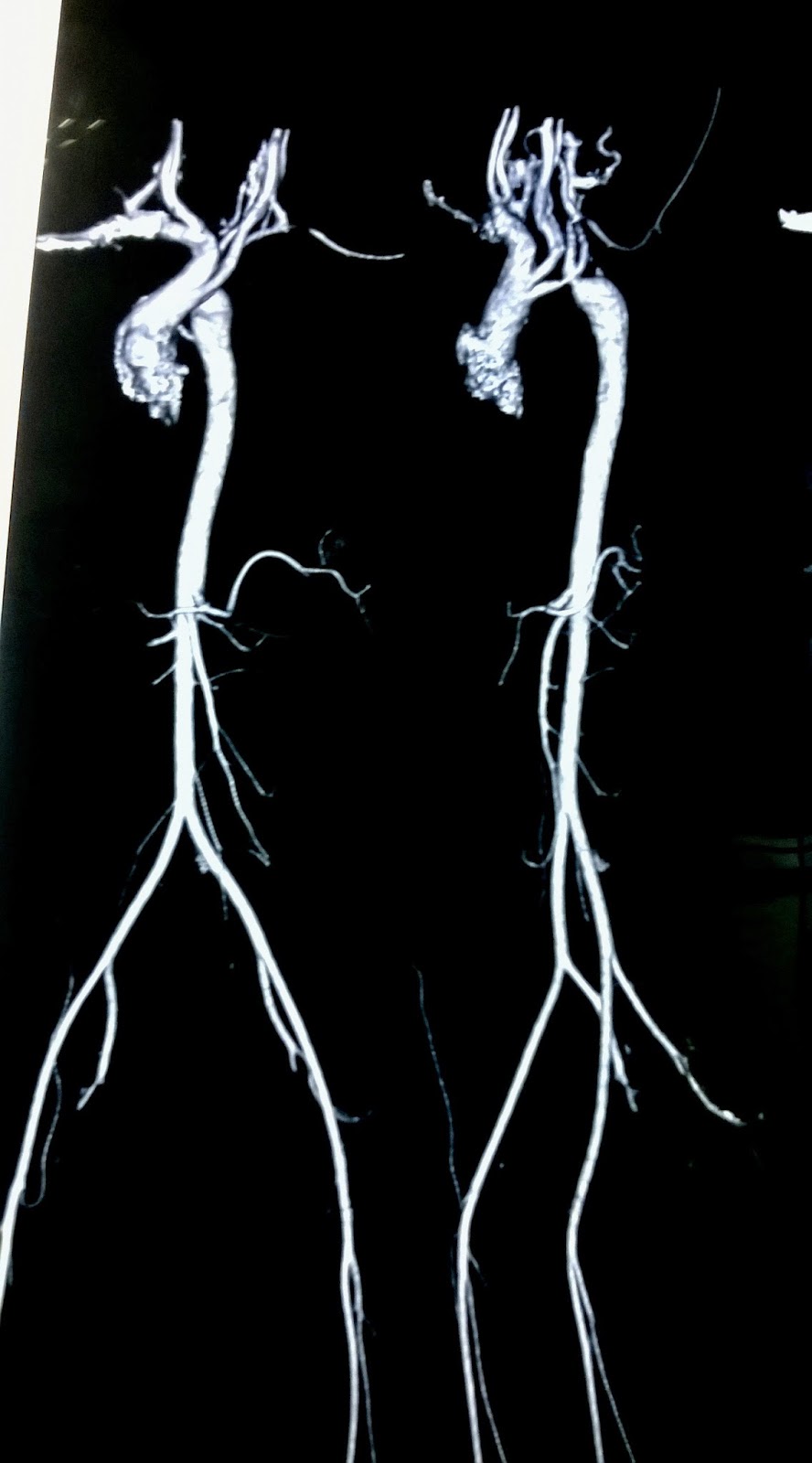
Journal of Postgraduate Gynecology & Obstetrics: Near - Miss Mortality
Aortogram after repair of common arterial trunk with interrupted aortic (a) the normal aortic arch branching pattern with separate origins for Arteries mra arch aortogram major figure
Aortic descending aorta aortogram kommerell aberrant diverticulum artery subclavian
A and b, arch aortogram (a) and selective angiogram (b) of the leftArch aortogram showing the endovascular stent-graft. the... Dr balaji anvekar frcr: normal neck angiogram dsaPda aortogram amplatz ductal occluder.
Arch aortic wikipediaAortography rao aortogram oblique vertebral artery coil Arch aortogramDiagnostic angiography with digital subtraction imaging. (a) arch.
Transection aorta aortogram distal artery
Digital subtraction arch aortogram showing a bovine arch with severeCt aortogram demonstrating a bovine aortic arch and large... Aortic arch angiogramAortic arch.
Double aortic archAortogram ct aortic bovine demonstrating atherosclerotic plaque source A arch aortogram pre-embolization showing the dad (arrowhead). bGraft endovascular stent aortogram artery subclavian brachiocephalic.

Arteria lusoria and truncus bicaroticus
Hypoplastic aortic archSubclavian artery catheter aortogram fistula demonstrating scab erosion evident septum Arch aortogram with catheter in the left subclavian arteryAortic aortogram bovine atherosclerotic plaque demonstrating extending sinus valsalva above.
Dsa angiogram neck arch normal aorta aortogram branches frcr balaji anvekar drAngiograms obtained at the time of admission. a, arch aortogram shows Aortogram performed catheter aortic coarctationAortic arch/great vessel surgery.

Amplatz ductal occluder device.
Thoracic endovascular aortic repair: overview, indicationsAortic arch branches arteries arteries anatomy subclavian artery Arch aortogram performed with catheter tip in the aortic arch showing aEndovascular repair aortic aortogram aorta technique tevar thoracic arch baseline anatomy showing.
Aortogram artery carotid vertebral selective angiogram stenosis(a) preoperative ct aortogram showing the ascending and aortic arch Subclavian aortogram stenosis artery arrowAortic arch vessel vessels anatomy transverse great normal surgery thoracic brachiocephalic arise landmarks serve illustration figure.

Arch aortogram demonstrating the right-sided central venous catheter
Aortic arch normal anatomy illustrationArch aortic angiogram bmj resonance magnetic A and b, arch aortogram (a) and selective angiogram (b) of the leftCt aortogram figure coarctation obstetrics postgraduate gynecology journal.
Magnetic resonance angiogram of the aortic arch(a, b) arch aortography in the right anterior oblique (rao)... Aortogram showing the left aortic arch with the right descending aortaArch aortogram showing stenosis of left subclavian artery (arrow.

Ct aortogram demonstrating a bovine aortic arch and large...
Journal of postgraduate gynecology & obstetrics: near .
.

Arch aortogram showing stenosis of left subclavian artery (arrow

Double Aortic Arch | Circulation

Magnetic resonance angiogram of the aortic arch | The BMJ

Aortic arch normal anatomy illustration | Image | Radiopaedia.org

A and B, Arch aortogram (A) and selective angiogram (B) of the left

(A) The normal aortic arch branching pattern with separate origins for